Pages

Showing posts with label Liver Cells. Show all posts

Wednesday, 20 March 2019

Milk Thistle Promotes Liver Detoxification

Liver deaths have jumped 65% in recent years because of an increase in non-alcoholic fatty liver disease largely due to obesity. Standardized milk thistle extracts have demonstrated remarkable protective and restoration effects on liver health.

January 2019
By Shirley Dannon
The liver is a primary defense against daily toxins.
People used to think that the main threat to their liver was alcohol.
Today’s nemesis is obesity. The result is an epidemic crisis of non-alcoholic fatty liver disease (NAFLD).1
Liver-related deaths jumped 65% between 1999 and 2016.2,3
Milk thistle extract has demonstrated benefits for non-alcoholic fatty liver disease, alcoholic liver disease, and cirrhosis.4-7
When standardized extracts of milk thistle are combined with a phospholipid delivery system, absorption is significantly enhanced.
The public tends to assume that liver disease affects only those who have abused alcohol.
What few people know is that as many as 30% of all adults suffer from non-alcoholic fatty liver disease, mostly caused by obesity.
There has been an almost two-fold increase in non-alcoholic fatty liver disease since 19911
Those whose diets consist of junk food and drinking sodas are falling victim to non-alcoholic fatty liver disease at alarming rates.8
The liver is one of the largest and most important organs in the body. It is susceptible to many sources of damage. These can include fatty liver disease infections, metabolic insults, and toxic poisonings including alcohol abuse.
Milk thistle extract has been shown to guard the liver from damage and reduce liver-related mortality.4,9-11
WHAT YOU NEED TO KNOW

Milk Thistle Extract and Liver Protection

  • The liver is one of the largest organs in the body and is critical for numerous functions, including removing environmental and dietary toxins from the body.
  • Liver disease is on the rise, affecting a large percentage of the population.
  • Extracts of milk thistle contain a powerful blend of liver-protecting compounds that help defend against toxins and oxidative stress.
  • Studies have demonstrated the ability of milk thistle extracts to protect the liver from a variety of causes of injury.
  • Standardized extracts of milk thistle complexed to phospholipids provide the greatest absorption into body tissues.

Factors behind Liver Disease

3D image of Liver
Poor diets, obesity, and other factors can lead to permanent liver damage. Chronic liver injury increases the risk for liver cancercirrhosis, and liver failure.
Here are some underlying correctable risk factors of the surging liver disease epidemic:
  • Excess alcohol ingestion (especially without accompanying it with antioxidants like n-acetyl-cysteine).12-14
  • Excess use of acetaminophen pain-relieving drugs (sometimes sold as Tylenol®).15-17
  • Abdominal obesity and excess body weight that contributes to nonalcoholic fatty liver disease.18 Almost 40% of adults are clinically obese according to the Center for Disease Control and Prevention (CDC).1
Considering the critical role of the liver in human health, and the growing incidence of liver disease, researchers have focused on the capacity of milk thistle extract to inhibit the most common causes and pathologies of liver damage.

The Potent Liver-Protection Effects of Milk Thistle Extract

Milk Thistle Flower
Extracts of milk thistle contain several natural compounds that demonstrate the ability to defend the liver from injury. These compounds offer multiple mechanisms of liver protection, including strong oxidant protection and anti-inflammatory properties.19,20
Here are some brief descriptions of studies looking at how milk thistle extract defends against some of the main drivers of liver disease.
PHOSPHOLIPIDS: IMPROVING THE BIOAVAILABILITY OF MILK THISTLE EXTRACT
Novel formulations of milk thistle extract have been developed that greatly improve the ability of the digestive system to absorb its beneficial compounds.
By joining these compounds with natural phospholipids, such as phosphatidylcholine, the bioavailability of standardized extracts of milk thistle is greatly increased.37
This new delivery system has been shown both to increase entry of milk thistle’s essential liver-protective compounds into the body and to maximize its beneficial effects.37

Non-Alcoholic Fatty Liver Disease

Non-alcoholic fatty liver disease is characterized by the deposition of fatty compounds in the liver. As the name implies, it is not caused by alcohol abuse and can occur even in individuals with little or no alcohol intake.
The prevalence of this disease is increasing, closely following the increases seen in related disorders, such as diabetes, metabolic syndrome, and obesity.1 In fact, non-alcoholic fatty liver disease can occur in up to 90% of obese individuals. 21 Fatty liver disease is on track to soon surpass alcoholism and viral hepatitis as the most common cause of chronic liver disease.
Early in its course, fatty liver disease may go unnoticed. However, it can progress over time to cause significant fibrosis of the liver and ultimately result in liver cancer, cirrhosis, and/or liver failure.
Scientists have demonstrated that, in preclinical models, extracts of milk thistle are capable of changing the abnormal metabolism associated with non-alcoholic fatty liver disease, reducing the accumulation of fatty substances and protecting cells from oxidative damage and death. 22-24
The mechanism of this protection may involve altering gene expression related to lipid metabolism, mitochondrial function, and antioxidant mechanisms.
In multiple studies of humans with fatty liver disease, intake of milk thistle extract has been associated with a reduction in fat deposition and improved enzyme blood markers of liver damage. 25-27
A meta-analysis of eight trials concluded that milk thistle extract effectively lowered liver enzymes, compared to controls, in non-alcoholic fatty liver disease patients.28
In randomized controlled trials, non-alcoholic fatty liver disease subjects treated with milk thistle extract for 48 weeks had markedly less fibrosis than those who received placebo.25,26,29

Alcohol and Other Liver Toxins

The liver is susceptible to an array of dangerous toxins known as hepatotoxins, including Amanita mushrooms, acetaminophen, arsenic, alcohol, and others.5,30,31
Research shows that milk thistle extract prevents liver damage when given before or during the exposure to a liver toxin.9,10,32,33
A common toxic cause of liver injury is alcohol abuse, and it appears to be on the rise, particularly in young adults.2,3 The best prevention of alcoholic liver disease is cessation of alcohol intake, but clinical trials have shown that milk thistle extract can even benefit people who’ve already developed the condition.6,11
One of these trials evaluated three different dose levels and found that the highest doses, 360 mg per day of the milk thistle extract preparation, resulted in the greatest improvement in liver enzyme levels.6 Another study showed that a group of patients with alcohol-induced liver disease who were treated with milk thistle extract showed improvements in liver enzymes and liver pathology.7
New studies show even what some people consider moderate alcohol ingestion can inflict an injurious impact.34 One might consider supplementing with milk thistle extract, n-acetyl-cysteine, vitamins B1, and C before ingesting ethanol.35
LIVER ENZYMES AS MARKERS OF LIVER INJURY
Measurement of liver enzyme levels is an important tool that clinicians and researchers use to identify ongoing liver injury.
These proteins, including AST and ALT, are normally present inside liver cells. However, when any pathology causes damage to liver cells, the levels of these enzymes rises in the blood, detectable on a simple blood test.
Elevated liver enzyme levels are therefore a useful marker of liver disease and can help to track response to treatment.
Milk thistle extracts have been found to decrease liver enzyme levels in many clinical studies, indicating protection from various types of liver injury.6,28,38-41

Cirrhosis

Man holding glass of alcohol
Cirrhosis is the end-stage of chronic liver injury. It can occur following years of ongoing liver injury from a number of possible causes, including viral and alcoholic hepatitis.
Cirrhosis is generally considered irreversible. But scientists have observed promising improvements with milk thistle extract in clinical trials.
In one such trial of 60 patients with alcoholic liver cirrhosis, those randomized to receive 450 mg of a milk thistle extract daily benefited from a reduction in a marker of liver fibrosis, along with improvements in oxidant reduction.36
Also, in a study of alcoholic and non-alcoholic patients with cirrhosis, 420 mg of milk thistle extract per day was associated with a higher overall four-year survival. This study showed that 58% of subjects supplementing with milk thistle survived compared to only 39% survival in controls (who did not receive milk thistle extract).4The greatest clinical improvement was observed in alcoholics.

Summary

Liver Enzyme Test
Among its myriad functions, the liver is crucial for detoxification, purifying the body of various toxic substances. As it performs these functions, the liver is itself prone to injury.
Acting through multiple mechanisms, compounds in milk thistle extract have the ability to protect the liver against toxins and metabolic disorders.
Health-conscious individuals can boost their body’s absorption of these protective compounds by taking standardized extracts of milk thistle that have been made more bioavailable via a phospholipid delivery system.

Tuesday, 28 March 2017

Liver Cells Grown From Human Skin Cells

Scientists at The Medical College of Wisconsin in Milwaukee have successfully produced liver cells from patients’ skin cells opening the possibility of treating a wide range of diseases that affect liver function.

By Medical College of Wisconsin in Milwaukee 
Oct 9, 2009 - 12:59:26 PM

Could Lead to Treatment of Liver Diseases


(HealthNewsDigset.com) - Scientists at The Medical College of Wisconsin in Milwaukee have successfully produced liver cells from patients’ skin cells opening the possibility of treating a wide range of diseases that affect liver function. The study was led by Stephen A. Duncan, D. Phil., Marcus Professor in Human and Molecular Genetics, and professor of cell biology, neurobiology and anatomy, along with postdoctoral fellow Karim Si-Tayeb, Ph.D., and graduate student Ms. Fallon Noto.


“This is a crucial step forward towards developing therapies that can potentially replace the need for scarce liver transplants, currently the only treatment for most advanced liver disease,” says Dr. Duncan.


Liver disease is the fourth leading cause of death among middle aged adults in the United States. Loss of liver function can be caused by several factors, including genetic mutations, infections with hepatitis viruses, by excessive alcohol consumption, or chronic use of some prescription drugs. When liver function goes awry it can result in a wide variety of disorders including diabetes and atherosclerosis and in many cases is fatal.


The Medical College research team generated patient–specific liver cells by first repeating the work of James Thomson and colleagues at University of Wisconsin-Madison who showed that skin cells can be reprogrammed to become cells that resemble embryonic stem cells. They then tricked the skin–derived pluripotent stem cells into forming liver cells by mimicking the normal processes through which liver cells are made during embryonic development. Pluripotent stem cells are so named because of their capacity to develop into any one of the more than 200 cell types in the human body.


At the end of this process, the researchers found that they were able to very easily produce large numbers of relatively pure liver cells in laboratory culture dishes. “We were excited to discover that the liver cells produced from human skin cells were able to perform many of the activities associated with healthy adult liver function and that the cells could be injected into mouse livers where they integrated and were capable of making human liver proteins,” says Dr. Duncan.


Several studies have shown that liver cells generated from embryonic stem cells could potentially be used for therapy. However, the possible use of such cells is limited by ethical considerations associated with the generation of embryonic stem cells from preimplantation embryos and the fact that embryonic stem cells do not have the same genetic make-up as the patient.


Although the investigations are still at an early stage the researchers believe that the reprogrammed skin cells could be used to investigate and potentially treat metabolic liver disease. The liver may be particularly suitable for stem-cell based therapies because it has a remarkable capacity to regenerate. It is interesting to note that the regenerative nature of the liver was referenced in the ancient Greek tale of Prometheus. When Prometheus was caught stealing the gift of fire from Zeus, he was punished by having his liver eaten daily by an eagle. This provided the eagle with an everlasting meal because each night the liver of Prometheus would re-grow.


The liver is a central regulator of the body’s metabolism and is responsible for controlling sugar and cholesterol levels, secretion of a variety of hormones, production of blood clotting factors, and has an essential role in preventing toxins from damaging other organs in the body.


It is possible that in the future a small piece of skin from a patient with loss of liver function could be used to produce healthy liver cells, replacing the diseased liver with normal tissue.


Recently, the National Institutes of Health’s National Institute of Diabetes and Digestive and Kidney Diseases through the American Recovery and Reinvestment Act have provided Medical College researchers, in collaboration with Markus Grompe, M.D., at the Oregon Health and Science University, a $1 million research grant to pursue the possibility of using reprogrammed skin cells to study and treat metabolic liver disease. Using this support, as well as donations from individuals throughout Milwaukee, Medical College researchers are currently producing reprogrammed cells from patients suffering from diabetes, hyperlipidemia, and hypercholesterolemia in an effort to identify new treatments for these diseases.


www.mcw.edu


http://www.healthnewsdigest.com/news/Disease_420/Liver_Cells_Grown_From_Human_Skin_Cells.shtml

Tuesday, 22 October 2013

Caffeine: the New Treatment of NAFLD?

Caffeine: the New Treatment of Nonalcoholic Fatty Liver Disease?


Unless your physician has specifically banned caffeine, there is no reason to shy away from a cup or two—or according to the latest research four—of coffee or tea.

Researchers from Duke-NUS Graduate Medical School and Duke University School of Medicine have revealed another possible health benefit of caffeine…this time as a treatment of nonalcoholic fatty liver disease.

Research published in the September 2013 edition of Hepatology suggests that increased caffeine consumption may decrease fatty liver cells in individuals afflicted with nonalcoholic fatty liver disease (NAFLD).

caffeine

When NAFLD Strikes

Your liver is responsible for turning food and drink into usable energy and for removing toxins and poisons from your blood. Nonalcoholic fatty liver disease strikes when more than 5% to 10% of your liver succumbs to the buildup of extra fat in your liver cells. NAFLD is caused by obesity, diabetes, high cholesterol, high triglycerides, hasty weight loss and poor nutrition. When left unchecked NAFLD can cause the liver to swell, leading to scarring (cirrhosis), liver cancer and eventual liver failure. Aside from diet and exercise, there is no treatment for NAFLD.

Approximately one-third of Americans have NAFLD, which is especially prevalent among diabetics and the obese (70% in fact). There are often no noticeable symptoms, but keep an eye out for the following:
  • Fatigue
  • Weight loss
  • Weakness
  • Loss of appetite
  • Nausea
  • Stomach pain
  • Spider-like blood vessels
  • Jaundice
  • Edema
  • Itching
  • Disorientation

Caffeine and Fatty Liver Disease

Using cell cultures and mice, Duke University researchers found that caffeine activates the break down of lipids (fatty acids) stored in liver cells. The fatty liver of mice fed a high-fat diet decreased when caffeine intake increased. This discovery indicates that four cups of coffee or tea a day can significantly help stall the progression of fatty liver disease in humans.

Paul Yen, M.D., associate professor and research fellow of the Duke-NUS Graduate Medical School’s Cardiovascular and Metabolic Disorders Program in Singapore expresses the researchers’ excitement as to the possibilities:

“This is the first detailed study of the mechanism for caffeine action on lipids in liver and the results are very interesting. Coffee and tea are so commonly consumed and the notion that they may be therapeutic, especially since they have a reputation for being “bad” for health, is especially enlightening.”

Health Benefits of Coffee and Tea

The health benefits of coffee and tea are substantial. Research suggests that caffeine may protect brain cells and reduce the risk of dementia. Coffee and tea have been proven to stimulate the gallbladder, thereby reducing the risk of gallstones, and caffeine also causes blood vessels to constrict, which reduces headache pain. Coffee and tea have been cited as preventative aids in heart disease, not to mention their high concentration of antioxidants.

This study certainly doesn’t negate the risks involved with excessive consumption of caffeinated beverages, but when consumed in moderation, coffee and tea impart many nutritive benefits. So, enjoy that cup of joe!


http://undergroundhealthreporter.com/caffeine-the-new-treatment-of-nonalcoholic-fatty-liver-disease

Friday, 30 August 2013

Understanding The Liver and Cholesterol

 Date: 12/21/2009    Written by: Jon Barron
Cholesterol & Liver Functions | Natural Health Newsletter 

liver location
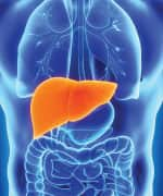
And now we come to the liver, one of my favorite organs. Certainly the heart, the brain, and the immune system get more play in the popular imagination than the liver, but that's only because the liver is so misunderstood. Next to the skin, the liver is the largest organ in the body. In many ways, it is the most important organ, and the last to be considered when it comes to health. In addition to being large, the liver is also a complicated organ involved in at least 200 separate functions.

Generally speaking, the liver performs a vital role in regulating, synthesizing, storing, secreting, transforming, and breaking down many different substances in the body. In this issue, we explore the anatomy and physiology of the liver in detail from a natural health perspective, and conclude with a discussion of how the body regulates cholesterol and why statin drugs may not be all that doctors promote.

Physical facts about the liver


As I mentioned above, the liver is the heaviest and largest gland inside the body, weighing in at about 3 pounds. Only your skin (also a single functioning organ) is larger. Your liver occupies almost the entire right upper quadrant of the abdominal cavity. (Remember that in virtually all medical diagrams, right and left are reversed.) It nestles up against the diaphragm on the top and against the ribs on the right -- stretching across the body, almost touching ribs on the left. Thus, barring extreme trauma such as bullet wounds and automobile accidents (or if it is not enlarged), it is fully protected -- a testament to how important the body considers the organ.

Physically, it is divided into four lobes, a large right and a small left lobe. Nestled between those two lobes are two less easily visible lobes, the quadrate lobe sitting on top and the caudate lobe sitting just underneath and extending to the bottom of the liver.

Obviously, a three pound organ cannot just "hang" in the abdominal cavity. It needs to be secured.  And in fact, it is suspended from the back of the diaphragm by two ligaments, the falciform and the suspensory ligaments. The falciform ligament in particular runs up through the entire liver, dividing the left and right lobes before attaching to the diaphragm. There is one other interesting note about the falciform ligament. The umbilical vein, when you are inside the womb, runs from the umbilical cord up between the left and right lobes of the liver. Within a week of birth, that vein is completely obliterated and replaced by the fibrous cord known as the falciform ligament.

The liver has a reserve capacity of some 50-80%. That means you can destroy up to 80 percent (and in some cases possibly even more) of the liver's function and have no demonstrable negative symptoms. And as amazing as that is, it's not the most amazing part. As I have mentioned frequently over the years when talking about detoxing the liver, the liver is one of the few human organs that can regenerate itself. It can actually regenerate (in a matter of weeks) up to an 80% loss of tissue.

Once regenerated, it will fill the same space it occupied before, and will take roughly the same shape as before. And when it's done regenerating, it stops! Though it grows faster than any cancer known to man, its regeneration does not become malignant, and the liver will stop growth at approximately its normal size. This is particularly useful after trauma such as an automobile accident that has damaged part of the liver. The damaged or diseased tissue can be removed by the surgeon with no loss of liver function, and in a matter of a few weeks, the liver will have regenerated all of its lost tissue. You've gotta love this stuff!

Your liver's blood supply


Before we begin discussing the liver's blood supply, which is unique in the body, it should be noted that everything in the liver begins with the three letters "hep", as in hepatic or hepatitis.

Your liver's vascular system


The right lobe of the liver is served by the right branch of the hepatic artery (a branch of the celiac trunk). The left branch serves the left lobe. Venous drainage occurs through the inferior vena cava, which cuts across the liver from top to bottom and receives venous drainage from the hepatic veins.

So far, nothing very interesting. But dig a little deeper and we find that the liver is unique in the entire body. In fact, the liver has an entirely separate circulation system to accommodate its special needs and functions. This is called the portal system.

Hepatic portal circulation


liver portal systemAs it turns out, all the physiology and pathology of the liver depend on this specialized circulation system. Functionally, the hepatic system is a venous system, ultimately returning used blood to the heart for reoxygenation. But unlike every other part of the venous system, it serves a second, even more important function. The portal system actually takes all of the veins that drain the organs of digestion and instead of returning their blood directly to the heart passes it through the liver.

Why?

Effectively, the portal venous system is responsible for directing blood from parts of the intestinal tract to the liver. All of the substances that you process and absorb in your small intestine must first travel to the liver for a final processing before continuing to the heart. In addition to the small intestine, the portal system also includes venous drainage from the spleen and pancreas.

So what is being processed in the liver?

Ultimately, we are talking about all of the protein, fat, and sugar molecules broken down in your digestive tract -- and all of the vitamins and antioxidants. Every nutrient you consume flows from the intestinal tract, through the portal system, and into the liver for processing and extraction. The liver thus plays a primary role in the digestive process. Specifically, the portal vein drains the inferior mesenteric vein, the superior mesenteric vein, the splenic vein, the gastric veins, and the esophageal veins. As you can see from the diagram, all of these veins dump into one vein, the inferior vena cava, just before it enters the lower part of the liver. From there it splits into many progressively smaller veins that ultimately reach every single cell of the liver before reversing the flow and reassembling, once again, as the inferior vena cava that exits through the top of the liver on its way back to the heart.

As a side note, many drugs that are absorbed through the intestinal tract are substantially metabolized by the liver before being parceled out for general circulation. This is the primary reason that so many drugs list liver damage as a notable side effect. On the flip side of the coin, processing by the liver "inactivates" some drugs, thus they cannot be taken orally. Nitroglycerin, for example, cannot be swallowed as it would be neutralized by the liver. Thus, it is taken under the tongue and absorbed sublingually, totally bypassing the portal system and the liver. Other drugs are administered through skin patches so they can be absorbed transdermally, once again bypassing the portal system and the liver.

As mentioned above, and as befits the special function of the portal system, the inferior vena cava does not continue as an uninterrupted thoroughfare through the liver. In fact, the portal system divides into a capillary bed of ever smaller venous capillaries in the liver sinusoids (see diagram below) formed by the cells of liver. It re-forms on the other side of the sinusoids as the hepatic capillaries and veins, which drain into the vena cava. Effectively, it is a venous-capillary-to venous-capillary system.

What happens inside the liver


Okay, we've laid out the location of the liver and the unique blood supply that supports it. Now let's talk about the structures inside the liver that do the actual work.
  • Liver lobules (aka hepatic lobules) are the microscopic working factories located inside the lobes of the liver.
  • Sinusoids are small swellings or "plates" between hepatocytes that act like small capillaries.
  • Hepatocytes are the liver cells that do all the actual work of the liver.


Liver lobules


The hepatic lobule is the structural unit of the liver. It consists of a roughly hexagonal arrangement of plates of liver cells radiating outward from a central vein in the center. Each lobule is approximately one to two millimeters in diameter, with tens of thousands of lobules per liver. At the vertices of the lobule are regularly distributed "portal triads," containing a bile duct and a terminal branch of the hepatic artery and portal vein.

liver lobule

The lobule is composed of multiple smaller units, called acini (which are just the grouping of cells at the end of each sinusoid) and is artificially defined into three zones, with Zone I closest to the portal vein and Zone 3 closest to the hepatic venule in the center of the lobule. Organization of the functional parts of the liver into lobules and acini between the portal vein and the hepatic vein allows for an easy exchange between the blood and the liver cells and a gradual filtration of blood as it moves through the sinusoids from Zone I to Zone 3. Contrary to blood flow, bile flows in the exact opposite direction, from Zone 3 to Zone I via a separate route to the portal bile ducts.

To summarize, nutrient-rich blood enters the liver via the hepatic artery and portal vein (remember, portal venous blood is rich in nutrients.) The blood from these two sources merges as it enters the sinusoids. Blood reaches the hepatocytes by detouring through capillaries at the sinusoids, where exchanges take place. The exchanges are made as the liver requires according to what zone the blood is in -- nutrients in, waste out, alcohol removed, etc. Blood then exits the lobule via the central hepatic vein, ultimately reentering circulation through the inferior vena cava that exits through the top of the liver. Note: the depleted blood returning from the legs and lower body is not involved in this exchange. It does not enter the liver. It is not altered in any way. Only the rich venous blood from the portal system is involved with the liver exchange before returning to the heart.

Sinusoids


Sinusoids are vascular channels lined by hepatocytes. As blood flows out of the hepatic arteries and portal veins, it flows through the sinusoids for "processing" by hepatocytes before it ultimately empties out through the central vein of each lobule, the hepatic venule, from where it ultimately makes its way to the heart. In addition to normal processing by hepatocytes, liver sinusoids are equipped with Kupffer cells that literally devour foreign pathogens such as bacteria that enter the sinusoids. As a side note, Kupffer cells are particularly vulnerable to damage from alcohol.

Hepatocytes - liver cells


Liver cells do the primary work in the liver -- everything from extracting oxygen and blood, to synthesizing cholesterol, to breaking down fats and sugars, to neutralizing toxins. That said, it's a little more complicated than that. In fact, although virtually all liver cells are fundamentally similar, their function varies according to their location in the liver lobule.  Zone 1 cells, for example, are located near the blood vessels that bring oxygen rich blood and nutrients into the lobule and are adept at oxidative liver functions such as cholesterol synthesis, the oxidation of fatty acids, glycolysis (the process that breaks down sugar for energy), gluconeogenesis (the formation of glucose), and lipogenesis. Zone 3 cells, on the other hand, specialize in detoxification.

What the liver cells actually do


The liver actually performs several hundred functions in the body. I can't cover them all in detail in this newsletter, but to summarize:
  • First, and probably foremost, there's protein synthesis. The liver synthesizes proteins from amino acids. It takes amino acids and assembles them as needed into complex proteins. It makes almost all prothrombin and fibrinogen (clotting factors), as well as albumin, the major blood protein. It also converts forms of amino acids from one to the other when needed for specific proteins.
  • It converts toxic ammonia (from amino acid conversions) into less toxic urea (which is excreted).
  • It uses amino acids and proteins for energy production or storage as fats and carbohydrates.
  • It metabolizes carbohydrates (CHO).
  • The liver is the storehouse of carbohydrates as glycogen (glycogenesis) and lipids (lipogenesis).
  • It can rapidly break down large amounts of CHO (glycogenesis) and release it as glucose into the bloodstream.
  • It can create glucose from lactic acid (gluconeogenesis).
  • It metabolizes lipids (fat).
  • The liver can store fats in various forms.
  • It can break down and release stored fat for extraordinary needs.
  • It cleanses the blood and discharges waste products.
  • The liver also excretes bilirubin, the broken-down pigments from dead red blood cells, by metabolizing it with bile salts and excreting it through the feces. Bilirubin is what makes our feces brown. If for some reason, bilirubin is not excreted (as in obstructive jaundice) the feces will turn clay-colored.
  • It neutralizes and destroys poisons and metabolizes alcohol.
  • The liver also detoxifies drugs and chemicals and virtually any toxin that enters the body. It excretes those toxins in two ways.
    • It neutralizes them and releases them into the blood, where they make their way to the kidneys and on out through the urine.
    • It dumps the toxins directly into the bile and, thence into the intestines for excretion.
  • It aids the digestive process by the production of bile, which is used for the breakdown of fats in the intestinal tract.
  • It helps the body resist infections by producing immune factors and by removing bacteria from the bloodstream.
  • It converts (conjugates) vitamin isolates as found in your vitamin pills into non-toxic forms your body can actually use -- and can then store some of those vitamins and minerals (iron and copper, for example) to be used as needed. In fact, the liver stores all the fat-soluble vitamins (A, B12, D, E, and K, for example). Water soluble vitamins such as vitamin C, on the other hand, are not stored in the liver and need to be taken daily, as any excess is expelled in the urine.
    • In addition, the liver is responsible for "activating" vitamin D so that your body can utilize it.
  • It helps maintain the body's hormonal balance.
  • It regenerates its own damaged tissue.
  • And it synthesizes cholesterol from fatty acids and removes excess cholesterol from the bloodstream as required.


Cholesterol

cholesterol molecule
In previous newsletters and in Lessons from the Miracle Doctors, I've covered the range of misinformation relating to cholesterol and heart disease. In this newsletter, I want to cover a different aspect of cholesterol -- how the body regulates it and, therefore, what we can do to optimize that process.

Cholesterol defined


Cholesterol is a fat soluble steroid, In fact, it is the most abundant steroid in the body. Far from being harmful, when properly regulated, it is a critically important molecule, essential in the formation of a number of key compounds, including:
  • Vitamin D
  • Progesterone
  • Estrogen
  • Testosterone
  • And adrenaline

It is also essential in the formation of every cell membrane in your body, not to mention the fact that your brain is mostly made up of cholesterol -- much of it in the myelin sheaths that insulate nerve cells and in the synapses that transmit nerve impulses.

As a fat soluble molecule, cholesterol cannot easily be carried in the blood -- a water based medium. Therefore, the body converts cholesterol into water-soluble molecules known as lipoproteins so it can be transported. Lipoproteins are composed of an outer shell made from a phospholipid which renders the particle soluble in water, a core of fats (lipids) including cholesterol, and a surface protein molecule (apolipoprotein) that allows tissues to recognize and take up the particle. Lipoproteins are characterized by their density: high density lipoprotein (HDL), low density lipoprotein (LDL), very low density lipoprotein (VLDL).

In simple terms, HDL lipoproteins are good for you. LDL lipoproteins "theoretically" promote arterial build up and are bad for you.

Where cholesterol comes from


cholesterol conceptDiet accounts for about 25% of the cholesterol levels in your body. Your liver synthesizes about the same amount, and the rest is synthesized in organs such as the intestines, the adrenals, and the reproductive organs. Obviously, trying to control cholesterol levels by solely changing diet will be effective only if levels are slightly out of whack. Although keep in mind, controlling diet is not just a question of regulating the cholesterol or fats that you eat; it is also a question of the soluble fiber (such as oat bran and psyllium husks) that you eat and which absorbs cholesterol and carries it out with your feces so that it never enters your bloodstream.

Unlike dietary changes, statin drugs seek to stop cholesterol formation in the liver by inhibiting a biochemical called 3-hydroxy-3-methylglutaryl coenzyme A reductase, which is required for cholesterol synthesis. Unfortunately, the reason for high cholesterol levels is only rarely due to over production in the liver; it is primarily the result of inadequate removal from the body. That means that statin drugs are not going to be the cause of the problem, but are addressing a symptom by artificially suppressing a properly functioning mechanism in the body. In scientific terms: that can't be good. We'll talk more about that later. But for now, let's talk about how your body actually regulates cholesterol levels.

As it turns out, your liver not only manufactures and secretes LDL cholesterol into the bloodstream; it also "down regulates" or removes LDL cholesterol from the bloodstream. In general terms, your liver oxidizes the cholesterol into a variety of bile acids which are then "pulled into the liver," carried into the bile ducts, and then on out through the intestines. As a side note, if the cholesterol becomes too concentrated in the bile and sits too long in the gallbladder, it can crystallize and form gallstones. (We'll talk more about gallstones in the next issue of the newsletter.) In any case, that's the general description of the cholesterol removal process. To understand exactly what's going on here, we need to examine it in a little more detail.

As it turns out, a healthy liver has a large number of active LDL receptor sites sitting on the surface of all the liver cells. When present and functioning properly, these receptor sites are associated with the rapid removal of LDL cholesterol from the blood -- and consequentially low blood LDL cholesterol levels. So why do these sites sometimes not perform as advertised?
  • Some people have an inherited genetic disorder (familial hypercholesterolemia) that causes them to be born with either a diminished number, or even zero, LDL receptors. For those people, statin drugs are a necessity. If the liver can never clear excess cholesterol, you have to stop production of cholesterol at its source.
  • Some people, on the other hand have normal receptor sites but as the result of bad diet literally clog up their livers so that the cholesterol cannot clear the receptors -- thus stopping the process of LDL removal. For these people, statin drugs are probably not the best "first" option.

How many of each are we talking about? The genetic condition affects maybe one in 500 people. For everybody else, we're talking about a self inflicted condition. In other words, the vast majority of cases of high LDL cholesterol are caused by a dietary inflicted blockage of the liver's LDL clearing mechanism. In fact, blockage occurs in two distinct places in the liver. First, fatty deposits can build up in the sinusoids, which prevents the bile from entering the bile ducts and physically clearing the liver -- essentially clogging the liver. Second, and more importantly, excess dietary fats can actually cause ingested cholesterol to build up in the membranes of liver cells, thus crushing the ability of those cells to process Sterol Regulatory Binding Protein (SREBP), which as its name implies, activates the gene in the LDL receptor site to tell it to take up cholesterol from the bloodstream. This literally stops the receptor sites from functioning as receptors -- totally shutting down the flow of cholesterol through the liver and on out through the bile ducts and colon. Depending on how many liver cells are blocked, this can lead to anything from a minor rise in cholesterol levels on up to a "your doctor is screaming at you" level. In any case, the use of statin drugs does nothing to change this underlying problem. They merely force your body to work around it.

cholesterol chart

Fortunately, there are options. If you can flush the excess fats and cholesterol that are unnaturally stored in the liver, your body's mechanism for regulating excess LDL cholesterol in the bloodstream will once again function properly -- automatically lowering your cholesterol levels. This is actually not that hard to do, although medical doctors have no idea how to accomplish it. It's called a liver detox/flush, and we will explore it in detail in the next newsletter.


Conclusion


Before we go, a couple of notes on statin drugs. I'm not very big of them for a number of reasons, including the fact that they have all kinds of side effects. But more to the point, they do nothing to address the underlying cause of high cholesterol levels for the vast majority of people who have the problem -- the clogging of your body's self regulating mechanism. Oh, and as a minor point, the connection between cholesterol and heart disease is not necessarily as automatic as you have been led to believe. But most significantly of all, one of the side effects of statin drugs is liver failure. Now, given the understanding you now have of what has caused the problem in the first place, how much sense does it make to take a drug that potentially destroys the one mechanism in your body that actually down-regulates cholesterol?

Absolutely none! The bottom line is that unless you are one of the 1 in 500 who has a genetic problem, statin drugs should only be used as a last resort. Far better to address the source of the problem -- which we will do next issue. Specifically, we will cover:
  • Liver function tests -- what they are and how to understand them
  • Things that can go wrong with the liver
  • The biliary tree, with emphasis on the gallbladder
  • The liver/gallbladder flush

http://www.jonbarron.org/article/understanding-liver-and-cholesterol

Friday, 7 December 2012

The Effects of Milk Thistle on the Liver

Dec 1, 2009 | By Tricia Mangan

The active ingredient in milk thistle is silymarin, a compound that is known to protect the liver. Silymarin acts to stabilize liver cell membranes, helping to repair and protect them from toxins and viruses, as well as assisting them to regenerate. Milk thistle has been used to prevent cancer and to treat alcoholism, cirrhosis, hepatitis, drug-induced liver toxicity, liver disease, food poisoning and indigestion. The effects of milk thistle on the liver are described below.
 

Improves Overall Liver Function

 
According to the Agency for Healthcare Research and Quality's (AHRQ) summary of milk thistle's effects on the liver, the most frequent benefit of milk thistle appears to be on aspects of overall liver functioning, including improvements in some liver enzymes and proteins such as aminotransferases, albumin, bilirubin, and/or malondialdehyde. More research is needed to determine whether milk thistle is more effective for acute, chronic or different types of liver diseases and what length of treatment is appropriate.

Repairs Damaged Liver Cells

 
Milk thistle's active ingredient, silymarin, is made of a group of flavonoids (silibinin, silidianin and silicristin) that stimulate protein synthesis. This stimulates the regenerative properties of the liver by promoting the growth of new hepatocytes, suggesting it may be useful in repairing livers damaged by alcohol, food poisoning and other toxins.

In the AHRQ summary of research findings on milk thistle, there was some evidence of improved liver functioning among those with acute viral and chronic cases of hepatitis and those with alcoholism, demonstrating milk thistle's regenerative abilities. However, the studies showed both positive and negative findings on ultimate survival rates among participants. More research is needed to clarify how much repair can be done and for what types of conditions milk thistle can improve survival.
 

Reduces Inflammation

 
Studies have suggested that silymarin is an anti-inflammatory substance that regulates molecules called inflammatory mediators and reduces enlargement of the liver.Those with hepatitis and liver inflammation may benefit from milk thistle's anti-inflammatory properties.
 

Prevents Liver Cell Damage

 
Milk thistle has the ability to alter the structure of the outer cell layer of hepatocytes (liver cells) in a way that prevents liver toxins from invading them. It may therefore protect the liver and slow down the advancement of cirrhosis from alcohol and drug toxicity as well as hepatitis. Laboratory rat studies offer some evidence that milk thistle blocks the toxic properties of some drugs that are poisonous to the liver, and that they can interrupt the recirculation of toxins in the liver and stimulate healthy re-growth.

Silymarin is also a natural antioxidant, and there is some early evidence from laboratory studies with animals that it may also help to prevent cancer by protecting the liver cells from toxic, tumor-causing chemicals.
Last updated on: Dec 1, 2009

What Causes Fatty Liver?


 Apr 10, 2010 | By Tina M. St. John, M.D.


What Causes Fatty Liver?
Photo Credit wurstplatte image by Daniel Fuhr from Fotolia.com
Abnormal accumulation of fat in liver cells causes a condition called fatty liver or steatosis. Fatty liver is important because it can trigger an inflammatory reaction, which may cause liver scarring and possibly cirrhosis. Steatohepatitis is the term for fatty liver disease with inflammation. Many conditions can cause fatty liver; frequent causes include excessive alcohol consumption, obesity and a high-fat diet. Fatty liver not related to alcohol use is called nonalcoholic fatty liver disease or NAFLD. If the condition is causing liver inflammation, it is called nonalcoholic steatohepatitis or NASH.

Excessive Alcohol
 
Excessive alcohol consumption is a common cause of fatty liver disease. Alcohol impairs the liver's normal metabolism of fat causing fat deposits within liver cells. Alcohol-related fatty liver may progress to alcoholic hepatitis.

Obesity
 
Obesity is strongly associated with fatty liver disease. Excess dietary fat and increased fat from body stores can cause fat buildup within the liver. As the rate of obesity has increased, so too has the rate of nonalcoholic fatty liver disease. In a 2008 review article published in "Clinical Science," Drs. David Preiss and Naveed Sattar reported an estimated 20 percent to 30 percent of adults in developed nations have NAFLD.
 
Type II Diabetes
 
Type II diabetes is associated with an increased risk for steatosis. The risk is highest among type II diabetics who are overweight and have poor blood sugar control.
 
Hepatitis C Infection
 
Infection with the hepatitis C virus may contribute to the development of fatty liver disease. This is important because the coexistence of chronic hepatitis C infection and fatty liver can accelerate liver disease progression. In a 2010 article published in "Nutrition and Metabolism," Dr. Silvia Mirandola and her colleagues note that approximately 50 percent of people with chronic hepatitis C have steatosis.
 
Medications and Supplements
 
Certain medications and supplements may cause steatosis including amiodarone, methotrexate, tetracycline, diltiazem, tamoxifen, valproic acid, cortisone, prednisone, highly active antiretroviral therapy (for HIV), and high doses of vitamin A.
 
Protein Malnutrition
 
Severe dietary protein deficiency can cause fat accumulation in the liver. Anorexia nervosa, prolonged fasting and restrictive low-protein weight loss regimens may cause fatty liver disease via protein malnutrition.
 
Wilson's Disease
 
Wilson's disease is an inherited disorder characterized by the accumulation of copper in the body. Copper accumulates in the liver causing progressive damage. Fat also commonly infiltrates the liver of people with Wilson's disease.
 
Lipid Storage Disorders
 
Hereditary disorders that interfere with the normal storage of body fats often cause steatosis. Examples of such disorders include Niemann-Pick disease, Gaucher's disease, Weber-Christian syndrome and abetalipoproteinemia.
 
Pregnancy
 
Acute fatty liver is a rare complication of pregnancy. With this condition, fat accumulates in the liver during the late stages of pregnancy. The disorder varies in severity, but is generally progressive until the baby is delivered. Although rare, this condition can be life-threatening.
 

References
http://www.livestrong.com/article/104288-causes-fatty-liver/

Monday, 11 June 2012

Regenerating liver inside the Body


Dean of Invention: Re-Gen Revolution

Originaly posted on: 28 April 2012

.

Dean of Invention: Growing New Organs Inside the Body (Series 1 Episode 8)

A doctor at the University of Pittsburgh finds a way to regenerate the liver inside the body cavity, an unprecedented feat!

Unfortunately, the video above finished a bit too early. In the report which was on Discovery Science broadcast on 15 April 2012 in Asia, it went on to show that the liver cells deposited themselves in the lymph nodes where they started to grow into mini livers in the mice, and taking on liver function capabilities. Hence the potential of growing new organs inside the body.

This programme was first shown in the UK in late 2010 and has probably been shown in many parts of the world. Catch it if you get an opportunity when shown or repeated.

Update on 3 June 2011: The programme is to be shown again on Sunday 10th June 2012 at 2200 hours on Astro Discovery Science Channel (554 in Malaysia).


----------------------------------------------------------------------------------------------

Updated on 11th June 2012:

3 medical institutions were highlighted on the programme:

a) Wake Forest Institute for Re-Generative Medicine

Surgeon Anthony Atala talked about using own body cells for growing body parts outside the body. Cells are coated onto a 'scaffold' in layers to form the organ. Organs grown included the bladder, heart valve, ear and skin.

From Wikipedia, the free encyclopedia:


Wake Forest Institute for Regenerative Medicine is a research institute affiliated with the Wake Forest School of Medicine and located at Winston-Salem, North Carolina, United States. Regenerative medicine is "a practice that aims to refurbish diseased or damaged tissue using the body's own healthy cells."[1] The institute opened its doors in May 2006 in an urban research downtown park in a 189,000-square-foot (17,600 m2) research building. The facility is part of the Piedmont Triad Research Park.[2][3]

Anthony Atala, M.D., is the Director of the Institute. He and many of his team came to North Carolina from the Laboratory for Tissue Engineering and Cellular Therapeutics at the Children's Hospital Boston and Harvard Medical School. Notable achievements announced at Wake Forest Institute have been the first lab-grown organ, a bladder, the artificial urinary bladder to be implanted into a human.[4][5] and stem cells harvested from the amniotic fluid of pregnant women. These stems cells are pluripotent, meaning they can be manipulated to differentiate into various types of mature cells that make up nerve, muscle, bone, and other tissues while avoiding the problems of tumor formation and ethical concerns that are associated with embryonic stem cells.[6]

http://en.wikipedia.org/wiki/Wake_Forest_Institute_for_Regenerative_Medicine


The following links were found on the web regarding the above institute:

Talks

Anthony Atala: Printing a human kidney

TED2011 Filmed and Posted Mar 2011


601,012 Views

Surgeon Anthony Atala demonstrates an early-stage experiment that could someday solve the organ-donor problem: a 3D printer that uses living cells to output a transplantable kidney. Using similar technology, Dr. Atala's young patient Luke Massella received an engineered bladder 10 years ago; we meet him onstage.

Anthony Atala asks, "Can we grow organs instead of transplanting them?" His lab at the Wake Forest Institute for Regenerative Medicine is doing just that -- engineering over 30 tissues and whole organs. Full bio »


Talks | TED Partner Series

Anthony Atala on growing new organs

TEDMED2009 Filmed Oct 2009 Posted Jan 2010


702,778 Views

Anthony Atala's state-of-the-art lab grows human organs -- from muscles to blood vessels to bladders, and more. At TEDMED, he shows footage of his bio-engineers working with some of its sci-fi gizmos, including an oven-like bioreactor (preheat to 98.6 F) and a machine that "prints" human tissue.

Anthony Atala asks, "Can we grow organs instead of transplanting them?" His lab at the Wake Forest Institute for Regenerative Medicine is doing just that -- engineering over 30 tissues and whole organs. Full bio »

---------------------------------------------------------------------------------------------

b) Weill Cornell Medical College, New York

The programme showed how blood and oxygen were introduced into the capillaries of the body parts using the cotton candy concept, mentioned in the above video.


---------------------------------------------------------------------------------------------


c) McGowan Institute for Re-Generative Medicine, University of Pittsburgh

Eric Lagasse was interviewed in the programme about his research on finding a way to regenerate the liver inside the body cavity.

The liver cells injected into the abdominal cavity of mice settled in the lymph nodes. The lymph nodes are incubator sites for producing immune system cells. One in ten lymph nodes produced more liver cells which took over carrying out the liver functions.

Eric Lagasse bio link:
http://www.mirm.pitt.edu/people/bios/lagasse.asp

Eric Lagasse Laboratory link:
http://www.mirm.pitt.edu/lagasse/default.asp


--------------------------------------------------------------------------------------------